
Platelets or thrombocytes react to bleeding from blood vessel injury by clumping, thereby initiating a blood clot. Platelets have no cell nucleus. Platelets congregate around a wound creating a cap to stop blood flow out of the tissue (clotting). Platelets also contain cytokines and growth factors which can promote wound healing and regeneration of damaged tissues.
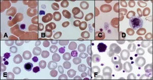
(A) Normal platelets; (B) and (C) agranular and hypogranular platelets in a patient with a myelodysplastic syndrome; (D) giant platelet; (E) platelet anisocytosis, large platelets and platelets with abnormal granulation in a patient with primary myelofibrosis; (F) platelet anisocytosis, a giant platelet and granulation anomalies in a patient with essential thrombocythaemia.
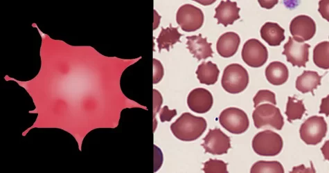
Infrequent acanthocytes are often encountered in hyposplenic conditions, but where they are very frequent this may indicate an uncommon or serious cause which needs to be communicated to clinicians. Light or severe liver disease causing coagulopathy and spur cell (acanthocytic) anaemia.
Primary Causes:
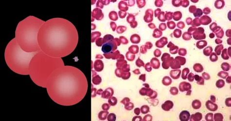
Agglutinates arise when antibodies attach to antigens on the membranes of adjacent red cells linking them together. The most common cause is “cold-reactive” IgM antibodies which do not cause overt symptoms. However, in some cases the effects may be clinically significant since antibodies may activate complement causing haemolysis, or the agglutinated cells can cause occlusion of small blood vessels in the cold (acrocyanosis). The clumped cells will sediment more rapidly leading to a raised erythrocyte sedimentation rate (ESR). Finally, the antibodies that cause cold agglutination may indicate an underlying malignancy (particularly lymphoma), or by acute infection.
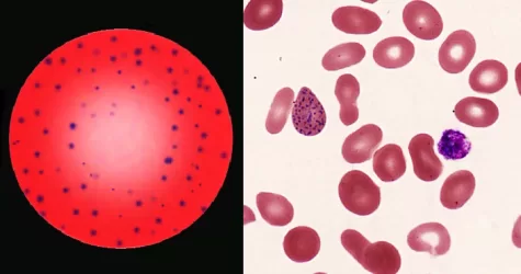
Most commonly arises when blood cell production is stressed or abnormal, may be associated with dysfunction of enzymes involved in RNA breakdown (either congenital deficiency or drug induced).
Normal individuals:
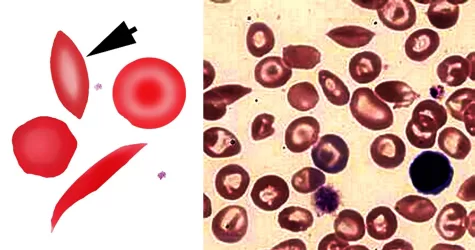
Although most closely associated with classical sickle disease (HbSS), boatshaped cells are also seen in compound heterozygotes between HbS and other abnormal haemoglobins.
Ignore boat cells in areas of blood movement, only consider if blood is static.
Inherited defects of haemoglobin with sickling tendency
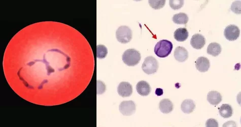
Cabot rings are ring-like or figure-of-eight loop-shaped inclusions composed of microtubule remnants from the mitotic spindle, or possibly nuclear remnants or abnormal histones. Can indicate B-12 anemia and related diseases, megaloblastic anemia, myelodysplastic syndrome, and lead poisoning.
Occur in states of stressed haematopoiesis:
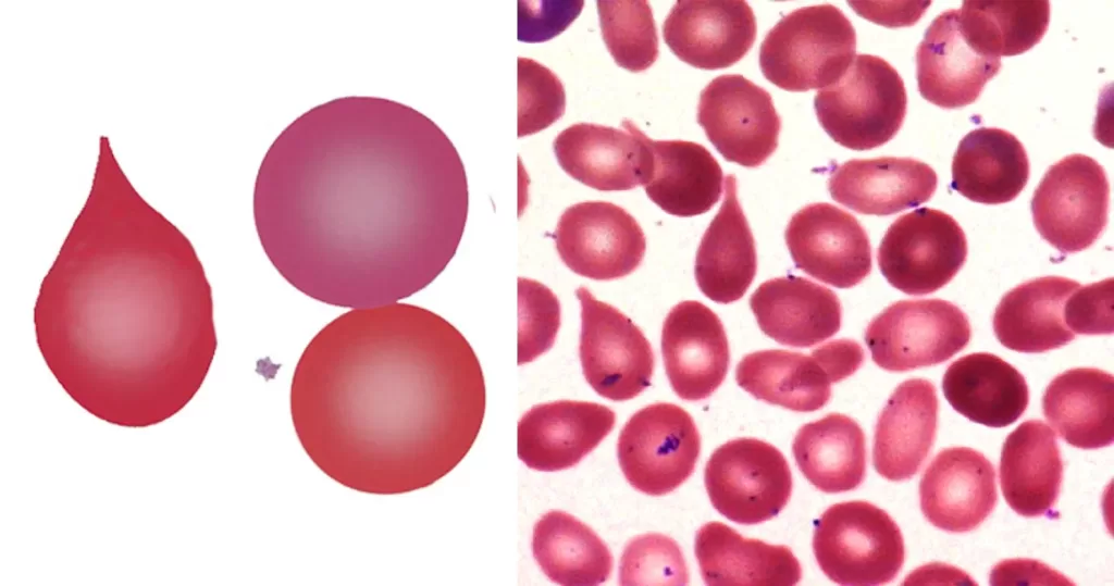
Strongly indicative of a “packed marrow”. May be the result of fibrosis (primary or secondary) or neoplasm (carcinoma or hematological neoplasm). May also arise where there is sustained or severe physiological increase in blood cell production (e.g. the expanded erythroid response to thalassemia). Less frequent tear drop forms may arise in other systemic disease. Ignore in fast-moving blood (vital force).
Intrinsic bone marrow disease
Iron deficiency or chronic disease. Multiple instances can indicate very severe hereditary pyropoikilocytosis.
Inherited defects
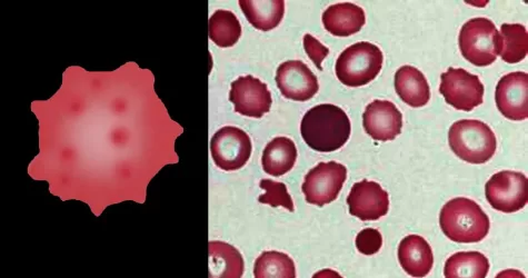
Always remember that echinocytes may be an artefact of blood storage or of cells on the edge of a live mount slide (artefactual echinocytosis), so look at the condition of other cells on the film and determine whether the echinocytosis is patchy in distribution. Where genuine there will usually be a significant systemic disease present. This most frequently will be renal failure.
Artefact
The presence of hypochromia indicates defective production of hemoglobin. Most cases result from iron deficiency or thalassemia – other typical features of these conditions should therefore be sought. Less frequently, hypochromia reflects defective iron utilization (e.g. chronic disease or sideroblastic anemia). The presence of hypochromia is not of itself an urgent problem unless there is severe anemia; however, it important to highlight the condition since clinicians may need to request further investigation to determine its cause.
Defective iron availability or usage
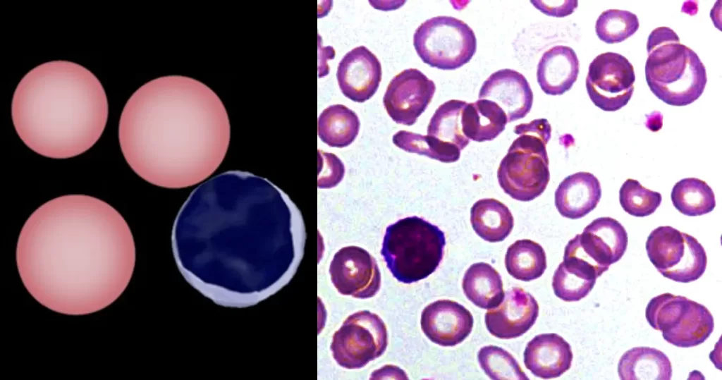
Howell-Jolly Bodies most commonly arise when spleen is absent or spleen function is impaired (hyposplenia). Occasional Howell Jolly bodies may arise in physiological conditions.
Hypo’splenism: Physiological
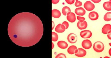
Implies damage to hemoglobin within the red cell often accompanied by cellular dehydration and membrane damage; acute oxidative damage to red cells should be considered.
Abnormal hemoglobin forms (look for associated typical cell forms)
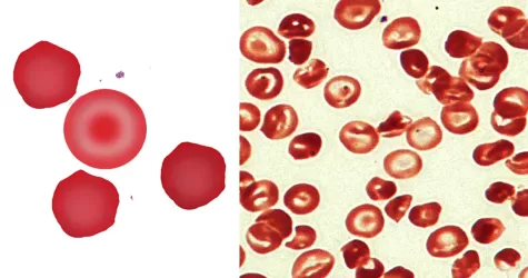
In some (although not all) cases, the pathological process may be life threatening particularly if they are associated with disseminated intravascular coagulation (DIC) or thrombotic thrombocytopenic purpura (TTP) knowledge of platelet count, clotting and additional morphological features such as fragments is essential.
Microvascular damage
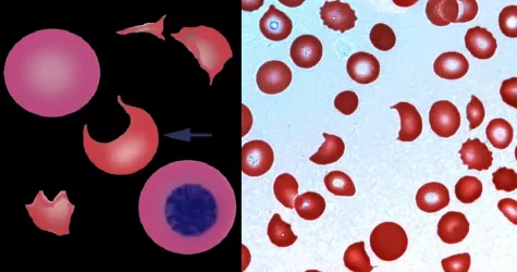
Morphological evidence of any accompanying disease should actively be sought. Most frequently these causes are B12 or folate deficiency, myelodysplasia, or liver disease.
Impaired cell division (nutritional or metabolic)
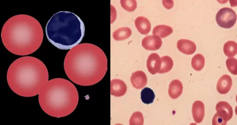
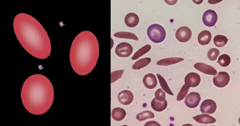
Myelodysplasia, iron deficiency, etc. S.E. Asian Ovalocytosis is a specific disorder that results from structural and functional defects of the band 3 protein causing ovalocytes with a stomatocytic appearance. May indicate previous malarial parasites.
Inherited Defects
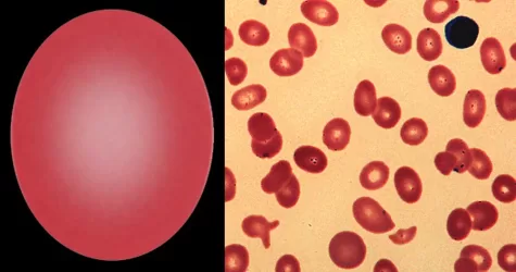
Small numbers of Pappenheimer Bodies may be seen in normal blood, particularly within polychromatic cells. When they are present in large number look for hyposplenic features, or for pathological states that have iron-loading or aberrant iron metabolism.
Normal Individuals
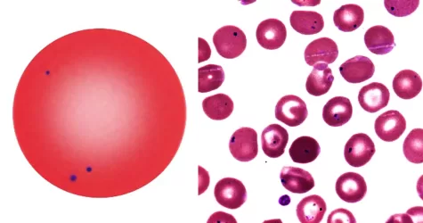
These cells are formed when sickle hemoglobin (HbS) is present together with hemoglobin C (HbC) to form a compound heterozygote disorder (HbSC disease)
Cause
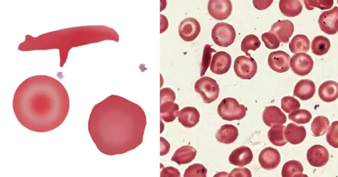
Fragmented cells are not found in normal blood. Sharp fragments may reflect “microangiopathic” damage – this form of fragmentation may therefore represent a medical emergency and should be reported immediately. More rounded fragments arise in significant dyserythropoiesis (such as severe myelodysplasia, membrane disorder or megaloblastic states), these are also important to diagnosis, but have a different origin.
Shearing processes (produce sharp fragments)
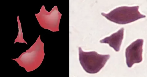
Indicates that the cells express the mutated gene for sickle hemoglobin (HbS), either in homozygous form (HbSS) or as a compound with another abnormal beta hemoglobin form. The number of these abnormal cells should not necessarily be considered an indicator of severity, but increased numbers of abnormal cells and polychromasia (or nucleated red cells) often occur during sickle crises.
Conditions with sickle hemoglobin
Sickle cell disease (HbSS)
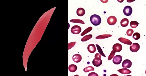
Spherocytes are found in all hemolytic anemias to some degree. Hereditary spherocytosis and autoimmune hemolytic anemia are characterized by having only spherocytes. Where spherocytes are very frequent autoimmune hemolysis or hereditary spherocytosis should be considered.
Inherited defects of membrane proteins (hereditary spherocytosis)
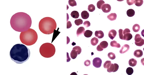
Severe membrane defects (e.g. hereditary pyropoikilocytosis). Toxin induced membrane damage: particularly Clostridium perfringens. Infrequent microspherocytes may appear as part of a spectrum of cells in many conditions with erythrocyte damage (e.g. fragmentation) or fragile production (e.g. megaloblastic states).
Inherited defects
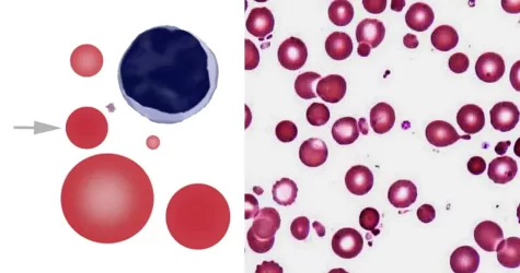
The area of pallor contains a central accumulation of hemoglobin giving the appearance of a “target”. Look for macrocytosis that may imply liver disease; or if MCV is normal or low consider a hemoglobinopathy (HbC, D or E).
Abnormal hemoglobin or abnormal hemoglobin synthesis
You can see how this popup was set up in our step-by-step guide: https://wppopupmaker.com/guides/auto-opening-announcement-popups/